Treatment for Skin Cancers and Skin Tumour Removals
The most common skin tumours in dogs are Mast Cell Tumours (MCT) (20%) and Soft Tissue Sarcoma (15%). Below are our quick guides to the diagnosis and treatment of each of these conditions. At Elizavet, we welcome referrals from first opinion practices and pet owners looking for treatment of skin cancers. Our collaborative approach deploys seamless communication, detailed case updates and shared care protocols to deliver good outcomes for every skin cancer referral. If you need more information, please contact us for a pre operative consultation with one of our veterinary surgeons, otherwise…
Mast Cell Tumour (MCT)
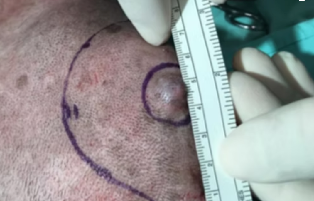
MCT tumours have different shapes; they can be on the skin (raised) or in the sub-cutis (under the skin), with or without hair, and may feel hard or soft to the touch. Even small tumours can release granules containing inflammatory mediators, which can in turn cause extensive skin irritation or inflammation around the tumour. This is called the ‘Darier Sign’.
Which breeds are most affected by mast cell tumours?
Certain breeds are predisposed to developing MCTs. The most affected breeds include Boxers, Boston Terriers, Pugs, Bull Terriers, Bullmastiffs, Cocker Spaniels, Staffordshire Bull Terriers, Fox Terriers, English Bulldogs, Dachshunds, Labrador Retrievers, Golden Retrievers, Beagles, Schnauzers, Chinese Shar-Peis, Rhodesian Ridgebacks, and Australian Cattle Dogs. (Shoop et al., 2015).
Diagnosis of MCTs
These types of tumours are frequently diagnosed via laboratory examination of samples collected through fine needle aspiration. Most of the time, this procedure does not require sedation; however, a skin biopsy under sedation and local anaesthesia may occasionally be needed for a final diagnosis.
MCTs are divided into two main categories: low-grade and high-grade tumours. High-grade tumours are more likely to spread within the body through the lymphatic system.
Ideally, a full blood and urine test should be performed to reduce anaesthetic risks. Fine needle aspiration of lymph nodes, abdominal radiographs, and ultrasound should be carried out to assess any internal spreading of the tumour. If internal metastatic lesions (spreading into the liver, spleen, or lymph nodes) are suspected, samples collected by fine needle aspiration from both organs should be examined.
Surgical removal of mast cell tumours
The surgical removal of the tumour can sometimes be challenging because of its anatomical position (e.g., on the limbs). Safe removal of the tumour is achieved by removing 2–3 cm of skin margin around the mass and a deep tissue plane (e.g., the fascia). The choice of 2–3 cm margins is based on laboratory results. Roots of high-grade tumours often spread within a 3 cm margin, so this distance is required for complete excision of the tumour.
Complete excision of the tumour and its roots will be confirmed through a laboratory test called histopathology examination. If a complete excision is not achieved, a review surgery, radiotherapy, or chemotherapy may be required.
Soft Tissue Sarcoma Removal (Skin)
Soft tissue sarcoma may arise from the skin or subcutaneous tissue. They are slow-growing and locally invasive tumours with a low metastatic rate (Tobias and Johnston, 2018). However, some high-grade tumours may spread through the bloodstream to the lungs.
Diagnosis of Soft Tissue Sarcomas
Fine needle examination can help rule out other types of tumours; however, it is not always diagnostic for soft tissue sarcoma. Ideally, the tumour should be examined through wedge or punch biopsy (histopathology). This often requires sedation and local anaesthesia.
Blood and urine tests are always recommended to reduce the risk of anaesthesia complications.
Although a CT scan is the gold standard for diagnosing lung metastasis, a chest radiographic examination is also advised before surgery. Three chest views with inflated lungs are sufficient to detect lung masses larger than 3 mm in diameter. Smaller masses can only be diagnosed by CT scan.
Removal of Soft Tissue Sarcomas
For complete excision of the tumour, 2–3 cm margins around the tumour and one deep fascia plane are always recommended (Tobias and Johnston, 2018). Complete excision of the tumour and its roots will be confirmed through histopathology examination. If incomplete removal occurs, review surgery, radiotherapy, or chemotherapy may be required.
If you need more information, please contact us for a pre operative consultation with one of our veterinary surgeons, otherwise…
Reference
Shoop J.W., Marlow S., Church D.B., English K., McGreevy P.D, Stell A.J., Thomson P.C., O’Neill D.G. and Bridbeltz D.C. 2015, Prevalence and risk factors for mast cell tumours in dogs in England [Online]: https://www.ncbi.nlm.nih.gov/pmc/articles/PMC4579370/