Hip Dysplasia and Total Hip Replacement
Hip dysplasia is a common orthopaedic condition in dogs and occasionally cats, caused by abnormal hip joint development that leads to looseness, pain and arthritis. As the joint deteriorates, pets may struggle to rise, show lameness, or become reluctant to exercise.
At our referral centre, we offer advanced diagnostics and tailored treatment to relieve pain and restore mobility. Early management focuses on physiotherapy, pain control, and weight management to slow joint degeneration. However, when arthritis or instability is severe, surgery provides the best long-term outcome. In this case Total Hip Replacement (THR) is the gold-standard treatment and for smaller pets or when THR isn’t suitable, a Femoral Head and Neck Ostectomy (FHO) can be an excellent alternative. If you are ready to make a referral request for the assessment and treatment of hip dysplasia please head over to our Make a referral page.
Diagnosis and medical management of Hip Dysplasia in dogs and cats
The information below covers the signs, diagnosis, medical management and surgical options for treating hip dysplasia. If you need more information before making a referral then please contact us to speak to one of our surgeons.
What are the first signs of hip dysplasia in dogs? The following are the most common clinical signs associated with canine hip dysplasia and degenerative joint disease:
- Stiffness, especially after periods of rest
- Exercise intolerance or fatigue following physical activity
- Lameness in one or both pelvic limbs (hind legs)
- A “bunny hopping” gait when running
- Difficulty standing up after exercise or prolonged lying down
- Reluctance to walk, run, jump, or climb stairs
In older dogs, chronic hip arthritis can lead to postural and muscular changes. These may include:
- Preference for sitting rather than standing
- Weakness and loss of muscle mass in the hind limbs
- Increased tone and size of the front limbs, as the dog shifts weight forward to compensate
Early recognition of these symptoms is crucial for preventing further joint damage and improving your dog’s quality of life.
-
- Photo 1
-
- Photo 2
-
- Photo 3
-
- Photo 4
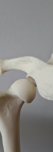
Photo 1: Normal Hip, The femoral head is sitting within the acetabulum.
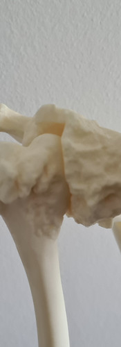
Photo 2: Joint laxity, the femoral head is not sitting within the acetabulum.
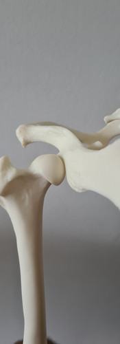
Photo 3: Joint laxity leads to dorsal subluxation of the femoral head.
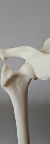
Photo 4: Developing of osteoarthritis and bone remodelling.
Signs of hip dysplasia in cats may include reduced jumping, reluctance to climb, stiffness after rest and subtle lameness. Cats often hide discomfort, so early veterinary assessment is recommended.
Accurate diagnosis of hip dysplasia in dogs is based on a combination of:
- Detailed clinical signs
- Physical orthopaedic examination
- Radiographic (X-ray) imaging
To confirm the diagnosis and determine the most effective treatment plan, a radiographic assessment is often performed under general anaesthesia. This ensures a clear view of the hip joint structure and allows for a thorough clinical evaluation.
If you suspect hip dysplasia or your pet is showing signs of joint pain, consult your vet for a full diagnostic workup and treatment recommendations.
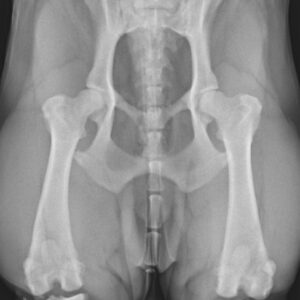
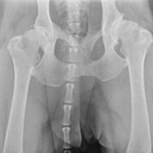
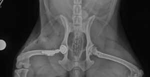
Photos: The image on the left shows a normal hip, while the image on the right shows severe arthritis and remodelling.
Medical management of hip dysplasia in dogs and cats focuses on improving mobility, reducing pain, and slowing joint degeneration through non-surgical approaches.
This conservative treatment plan typically includes:
- Weight management to maintain a lean body condition
- Physiotherapy to strengthen muscles and improve joint function
- Joint supplements such as glucosamine and chondroitin
- Pain control with NSAIDs and other appropriate medications
The primary goal of medical management is to enhance the pet’s quality of life by preserving mobility and comfort. While these therapies do not cure hip dysplasia, they play a critical role in delaying progression of osteoarthritis and managing chronic pain.
It is important to note that medical management is considered palliative, meaning it focuses on symptom relief rather than correcting the underlying joint abnormality.
Surgical treatment for hip dysplasia is recommended in cases where medical management is insufficient or when joint instability is advanced. The choice of surgery depends on the age of the pet, the severity of hip laxity and the presence of degenerative joint changes.
In young animals with minimal or no arthritis, early surgical interventions aim to correct hip joint alignment and reduce future degeneration:
- Double Pelvic Osteotomy (DPO): Rotates the pelvis to improve coverage of the femoral head by the acetabulum, reducing subluxation.
- Juvenile Pubic Symphysiodesis (JPS): A minimally invasive technique performed in puppies (under 16 weeks old) to alter pelvic growth and improve hip congruency.
These procedures are most effective before signs of arthritis develop, and they help slow the progression of hip dysplasia.
When hip dysplasia has progressed or joint instability is severe, especially in older pets or those with marked lameness, more advanced surgical options are considered:
- Total Hip Replacement (THR) — also known as dog hip replacement
- Femoral Head and Neck Ostectomy (FHO)
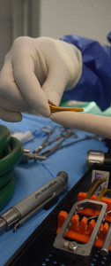
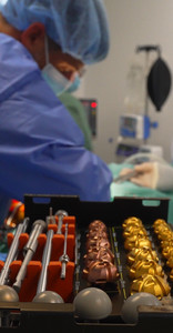
Surgical decision-making for hip dysplasia depends on:
- Your pet’s age and weight
- Extent of joint damage
- Your goals and expectations as an owner
- Your surgeon’s recommendation – this will be a joint decision
A thorough clinical evaluation and advanced imaging (X-rays or CT scans) help determine the most appropriate approach.
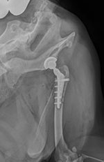
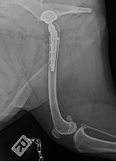
Total Hip Replacement (THR) surgery in dogs and cats
Total hip replacement (THR) is an advanced surgical procedure used to treat severe hip dysplasia and degenerative joint disease in dogs and cats. It involves the complete removal of the diseased hip joint and replacement with a prosthetic implant, restoring both function and comfort. This procedure is designed to; Eliminate chronic pain, restore full range of motion, improve quality of life and to enable pets to return to active, pain-free movement
We welcome referral requests for THR from pet owners and veterinary practice from across London and beyond.
THR involves the surgical removal of the femoral head and neck, which is then replaced with a prosthetic femoral head that fits into a newly implanted acetabular cup. These components replicate the anatomy of a healthy hip joint, offering a stable, smooth and pain-free range of motion.
At Elizavet, we are proud to offer Kyon Total Hip Replacement (THR) implants, a premium solution known for precision engineering and long-term durability. The key benefits of the Kyon THR system include…
- Implants made from high-grade titanium and ceramic, ensuring superior biocompatibility and reduced wear.
- Implants that achieve a precision fit to each patient, maximising stability and function.
- The system minimises risk of implants loosening or complications due to advanced fixation design.
By using Kyon implants, we ensure that every THR procedure delivers optimal outcomes in joint longevity, post-operative mobility, and patient comfort.
With proper case selection and post-operative care, THR can restore your pet’s full functional activity, including running, jumping and playing without pain. Most patients experience rapid improvements in comfort and mobility within weeks of surgery.
Consistent monitoring and timely veterinary intervention are imperative to identify and address potential complications, ensuring a favourable outcome for the THR procedure.
While the overall complication rate for canine THR is estimated at 5–8%, most complications are rare and manageable. These include:
- Re-luxation of the joint
- Infection
- Femoral fracture
- Sciatic nerve irritation
Of these, infection is the only complication that may require removal of the prosthesis. However, we take every precaution to minimise risk, including the use of iodine-impregnated drapes, Semini project meds and strict sterile procedures.
To provide added peace of mind, if selective complications* occur within one month post-surgery, we offer flat-rate coverage for any necessary interventions.
*Please ask for details.
See more about our THR treatments
instagram.com/p/DMDU1XHC5nb/
instagram.com/p/DCwU0qQiKmO/
Femoral Head and Neck Ostectomy (FHO) surgery for hip conditions
This surgical procedure is recommended for; Recurrent hip luxation (hip dislocation), Severe, non-repairable fractures of the acetabulum or femoral head and Advanced osteoarthritis that’s causing chronic hip pain. We welcome referral requests for FHO from pet owners and veterinary practice from across London and beyond.
The primary goal of FHO surgery is to eliminate pain caused by bone-on-bone contact in the hip joint, specifically between the femoral head and the acetabulum. Using an oscillating electric saw, the femoral head and neck are surgically removed. After removal, joint function relies on the surrounding soft tissues, tendons, and muscles, rather than the bony structures.
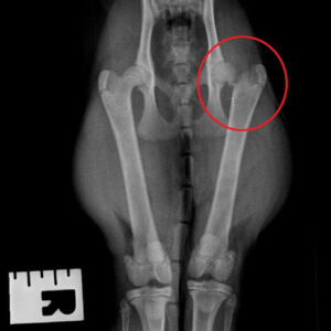
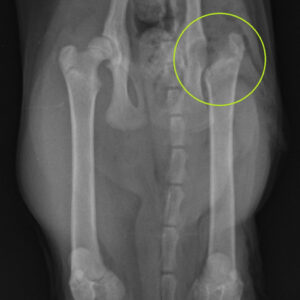
Photos: Fracture of the femoral neck (red circle) treated with femoral head and neck excision (green circle).
Effective pain management and precise surgical technique support early limb use and the development of strong muscle tone, essential for long-term joint stability. A structured rehabilitation programme, including canine hydrotherapy or physiotherapy, is recommended once the patient begins weight-bearing, to encourage mobility and support recovery.
Choosing between THR and FHO
When treating hip dysplasia in dogs and cats, the choice between Total Hip Replacement (THR) and Femoral Head Ostectomy (FHO) depends on the patient’s size, activity level, and overall health. THR is the gold-standard option, restoring the hip’s normal anatomy, joint stability, and near-complete function, making it ideal for larger or more active pets. However, when THR is not possible—due to cost, health risks, or anatomical limitations—FHO remains an excellent alternative. By removing the femoral head, it eliminates bone-on-bone pain and allows a false joint to form. This technique is particularly effective in smaller animals under 10 kg, providing reliable pain relief and mobility. If you’d like more information before making a referral then please contact us to talk to our surgical team.