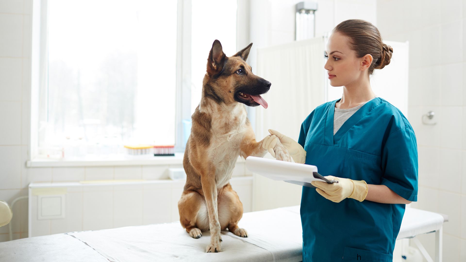
Elbow Dysplasia and Arthroscopy
Elbow dysplasia is a developmental orthopaedic condition causing pain, lameness, and reduced mobility in dogs — and occasionally cats. It occurs when the three bones forming the elbow joint (humerus, radius, and ulna) do not fit together correctly, leading to abnormal pressure, cartilage wear, and arthritis. Common in young, large-breed dogs such as Labradors, German Shepherds, and Golden Retrievers, signs include forelimb lameness, stiffness after rest, and reluctance to exercise.
Early diagnosis using X-rays, CT scans, or arthroscopy is key to slowing joint degeneration and improving outcomes. Treatment depends on severity and may include medical management — such as pain relief, physiotherapy, and weight control — or surgical options. Minimally invasive arthroscopy can remove fragments and treat damaged cartilage, while more advanced procedures like PAUL-2 osteotomy or Total Elbow Replacement address severe cases.
If you are ready to make a referral request for the assessment and treatment of elbow dysplasia, please …
Diagnosis and management of elbow dysplasia in dogs and cats
The information below covers the signs, diagnosis, medical management and surgical options for treating elbow dysplasia. If you need more information before making a referral, then please contact us to book a consultation with one of our surgeons.
Elbow dysplasia, a syndrome causing arthritis, pain, and limited joint movement, primarily affects young, large and giant breed dogs. Breeds such as German Shepherds, Golden Retrievers, and Labrador Retrievers are commonly affected, though smaller breeds like French Bulldogs can also develop the condition.
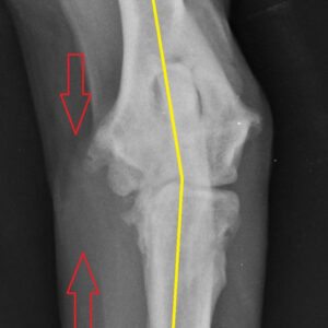
Typical indicators include forelimb lameness, stiffness after rest, reduced willingness to exercise and elbow joint pain or swelling. If you notice these signs you should seek veterinary assessment promptly.
Early intervention is crucial to minimise arthritis formation. Diagnosis involves radiographs, and in inconclusive cases a CT scan or arthroscopic exploration may be necessary. Early signs can be detected between 4 and 6 months of age. Early treatment, especially before visible soreness occurs, through corrective osteotomy can enhance joint congruency. This results in a milder progression of osteoarthritis and a better long‑term outcome.
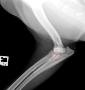
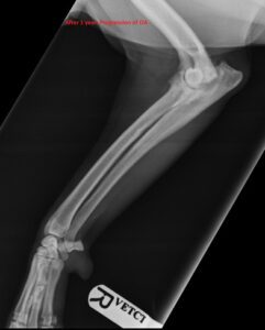
Treatment options for elbow dysplasia in dogs
Treatment options vary by clinical case but can be medical or surgical. Evidence‑based treatment paths for elbow dysplasia in dogs are selected after imaging and orthopaedic examination.
Medical treatment for elbow dysplasia in dogs
Medical treatment includes pain management, physiotherapy, weight control, and joint supplements. Some cases can be managed medically; however, surgery generally provides better outcomes by slowing arthritis progression and reducing pain.
Surgical treatment options for elbow dysplasia in dogs
A minimally invasive procedure using a small camera to inspect and treat cartilage damage and, if required, remove small bony fragments.
Used in severe cases, this procedure involves cutting the ulna bone and shifting body weight to alleviate lameness. Significant improvement is often observed, although some residual lameness may persist.
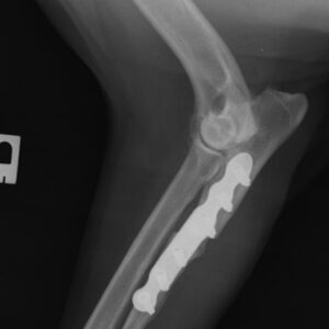
Reserved for end‑stage cases with severe osteoarthritis, this replaces the entire elbow joint with a metal prosthesis. Post‑operative infection is a notable complication and may lead to implant removal, elbow arthrodesis, or limb amputation.
Long‑Term management of elbow dysplasia in dogs
All patients with elbow dysplasia, whether surgically treated or not, require long‑term management of secondary arthritis. Early surgical intervention helps minimise arthritis progression and reduces long‑term pain.