Veterinary Ultrasound for Dogs & Cats
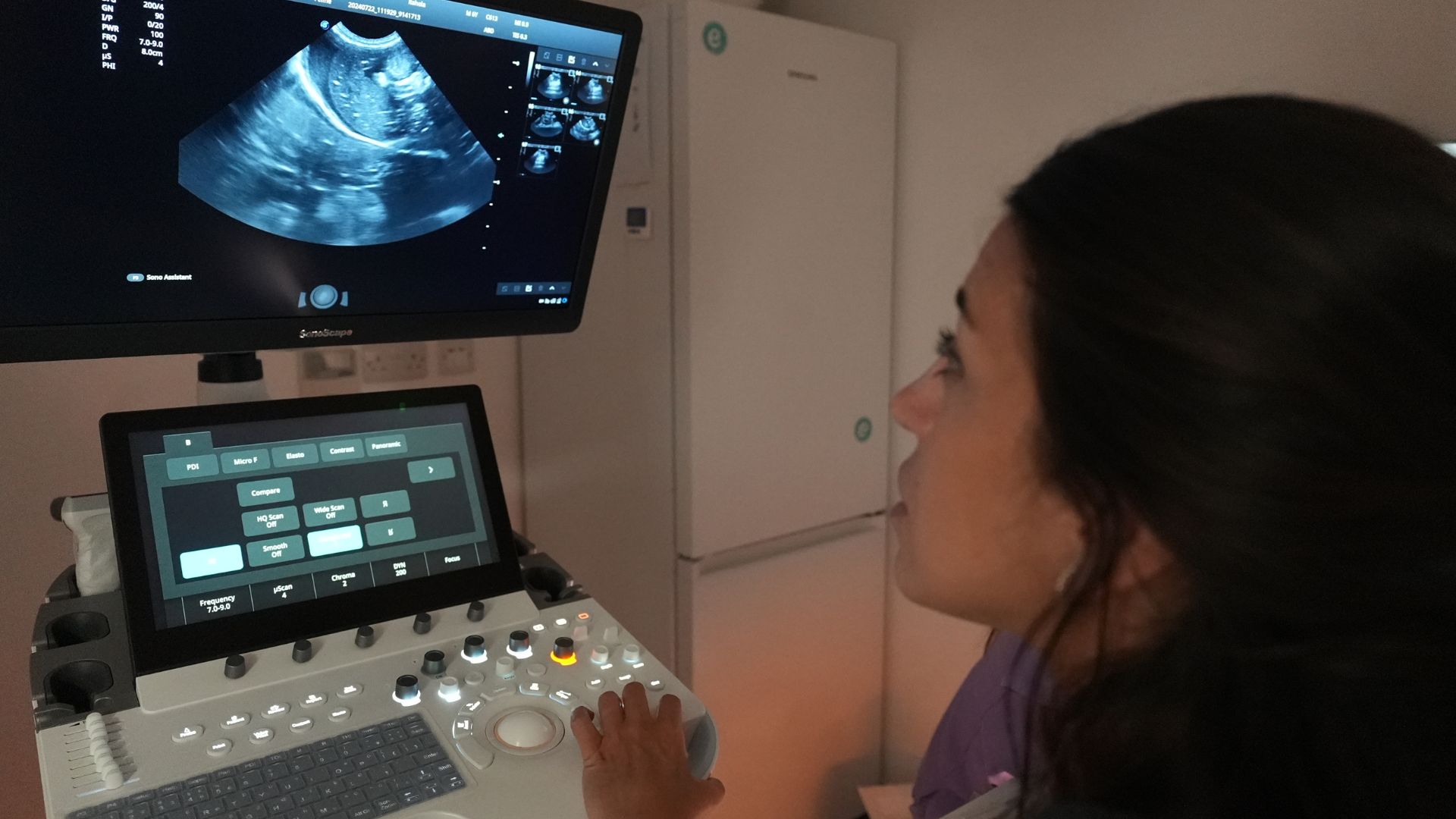
Veterinary ultrasound is a non-invasive diagnostic tool that enables veterinarians to visualise internal organs and tissues. Using high-frequency sound waves, it produces real-time images of the heart, liver, kidneys and other vital organs of dogs and cats. This safe and effective technique allows accurate diagnosis without the need for surgery.
Both veterinary practices and pet owners from our local area, around Camden and across London come to Elizavet for ultrasound diagnostics. If you need more information, please contact us to speak to one of our veterinary surgeons, otherwise …
An Introduction to our Ultrasound Facilities and Services
What is veterinary ultrasound used for?
Veterinary ultrasound is widely used to assess internal structures, monitor pregnancies and guide fine-needle biopsies. A dog or cat ultrasound check is typically performed quickly, with minimal restraint, making it a safe and low-stress procedure. This advanced imaging technique provides detailed insights, improving diagnostic accuracy and supporting effective treatment plans.
During the procedure, a small patch of fur is clipped from the area being examined, and a gel is applied to the skin to improve image clarity. The examination is pain-free, although mild sedation may occasionally be required to keep anxious animals relaxed.
Advanced ultrasound technology at Elizavet
At Elizavet, we use state-of-the-art equipment featuring multiple probes designed for specific organs. Our system includes Colour Doppler and elastography capabilities to deliver highly accurate imaging results. With advanced software and high-resolution displays, we ensure exceptional image quality. Our sonographers hold postgraduate qualifications in diagnostic imaging, ensuring expert interpretation and care.
Why ultrasound is used in veterinary medicine
Dog ultrasound and cat ultrasound are essential diagnostic tools in veterinary practice, allowing clear visualisation of soft tissues, organs, and blood flow without surgery. The main uses include:
Diagnostic imaging is used to assess a variety of organs and conditions including;
- Abdominal organs – it’s used to examine the liver, kidneys, bladder, spleen, and gastrointestinal tract for abnormalities such as tumours or inflammation.
• Pregnancy monitoring – it confirms pregnancy, estimates gestational age, and assesses foetal health.
• Cardiology – it evaluates the heart’s structure and function using echocardiography to detect heart disease.
In emergencies, point-of-care ultrasound (POCUS) enables rapid assessment of internal bleeding, fluid build-up, or organ rupture, helping veterinarians make quick, informed decisions for critical patients.
Ultrasound allows our vets to monitor disease progression, assess treatment response, and track recovery. For example, it is used to measure tumour shrinkage during cancer therapy or to monitor chronic organ conditions.
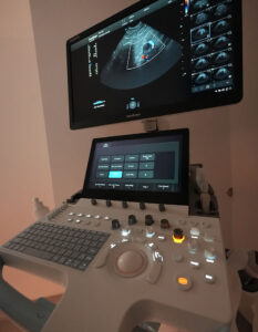
Colour Doppler ultrasound measures blood flow within vessels, offering valuable insights into cardiovascular health. It helps detect abnormalities such as blood clots, restricted flow, or heart valve issues.
Fine needle aspiration involves collecting small tissue samples for analysis under a microscope. When guided by ultrasound, it allows precise targeting, ensuring accuracy and minimising discomfort. This minimally invasive procedure is usually performed without sedation.
An ultrasound-guided core biopsy safely collects tissue samples from suspicious masses. It avoids surgical procedures while providing accurate diagnostic information to guide treatment decisions.
Echocardiography is a specialised ultrasound used to evaluate the heart. Symptoms such as coughing, fatigue, or fainting may indicate heart disease, though many pets show no signs until advanced stages. Certain breeds are predisposed to heart conditions, making regular screening important. If abnormalities are detected, your vet may recommend an echocardiogram for a precise diagnosis.
If you suspect your pet may have heart disease or if your veterinarian notices any irregularities, an echocardiogram performed by one of Elizavet’s veterinary cardiologists can provide a clear diagnosis. Speak with your vet to determine whether an echocardiogram is recommended for your pet’s health.
The cost of ultrasound cost – what to expect
The cost of an ultrasound examination depends on a number of factors including the complexity of the examination, the area being scanned and any additional diagnostics required. At Elizavet, we always provide transparent pricing and discuss all costs with you before proceeding with any procedures. If you’d like an estimate or more information, please contact us to speak to one of our diagnostic team, otherwise …